Indoor Mold's Real Threat Isn't the Stain — It's the Air Around It
A new method for detecting airborne mycotoxins confirms what the stain on your wall can't tell you: the toxic part already left.
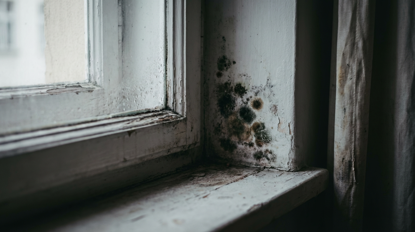
There is a dark smudge in the corner of the bathroom, or behind the wardrobe, or along the windowsill where condensation pools every winter. Maybe you have been meaning to clean it. Maybe you have been vaguely aware of it for months. What you probably have not been thinking about is the fact that it has been exhaling into the room the entire time, shedding chemical metabolites into the air you breathe while you sleep, while you eat breakfast, while you sit three feet away and watch television.
The visible mold colony is the part that bothers people. It looks alive because it is alive — a fungal network spreading across a damp surface, sending reproductive spores into the air, feeding on cellulose and wallpaper paste and the microscopic film of grease and dead skin that coats most indoor surfaces within days of cleaning. People scrub it off, paint over it, or simply avoid looking directly at it. But the biological activity of a mold colony is not limited to the part you can see, and the threat it poses to indoor air quality is not limited to the spores you might inhale. In 2025, a study published in Analytical and Bioanalytical Chemistry did something that had never been done cleanly before: it developed the first highly sensitive method to actually quantify airborne mycotoxins — the toxic secondary metabolites produced by mold — as they float freely through an occupied room. The results were not reassuring.
Mycotoxins are not the same as spores, and they are not the same as the visible mold itself. They are chemical weapons — small, stable, lipid-soluble molecules that fungi synthesize as byproducts of their metabolism, often as a way of defending territory against competing microbes. Aflatoxin, ochratoxin A, trichothecenes, zearalenone: these are the names food safety laboratories watch for in grain and nut supplies, where they can accumulate to regulatory-flagging concentrations. The assumption, for a long time, was that indoor air was a different problem. Spores, yes. Mycotoxins, maybe, but too dilute to quantify well, too hard to separate from background contamination. The new measurement method dismantled that assumption. The mycotoxins are there. They are measurable. And they are arriving in your lungs as particulate matter, attached to dust and fungal fragments too small to see and far too small to feel.
What makes this finding scientifically important is the word "passively." Mold does not need to be disturbed to release mycotoxins into the air. It does not require a renovation project, a vigorous scrubbing, or a burst pipe. A colony sitting quietly on a damp wall, doing nothing dramatic, continues to metabolize and continues to shed. The air in a moldy room is not static. It is a slow chemical conversation between the colony and everything that breathes in that space.
What Mold Is Actually Doing on Your Wall
A mold colony is not a stain. It is a living structure. What you see on a surface is the surface expression of a much denser hyphal network — a web of thread-like filaments called hyphae that have already penetrated the substrate beneath. On drywall, that means the fungus is inside the paper facing. On wood, it is inside the grain. On painted concrete, it is behind the paint layer, feeding on the organic binders. The dark or greenish or fuzzy surface is the reproductive layer: conidiophores bearing conidia, the spores the colony is broadcasting into your air to found new colonies elsewhere. By the time a colony is visible, it has typically been established for days to weeks. It has eaten, spread, and reproduced before it was noticed.
The most common indoor mold genera — Aspergillus, Penicillium, Cladosporium, and Stachybotrys — have different moisture requirements, different surface preferences, and different toxin profiles. Stachybotrys chartarum is the one that gets the most public attention because it produces trichothecene mycotoxins and favors persistently water-damaged materials, and it is genuinely concerning. But Aspergillus and Penicillium species are far more ubiquitous in ordinary indoor environments and are fully capable of producing ochratoxin A and a suite of other bioactive compounds. The dramatic "black mold" narrative that dominates popular coverage is misleading in both directions: it overweights one species while underweighting the toxin-producing potential of the molds that are actually common in most homes.
“The visible mold is the colony's face. The mycotoxins are its breath.”
The Measurement Problem That Hid the Risk
For years, indoor air quality research focused on airborne spore counts because spores were what could be captured and identified. They are relatively large, they have recognizable morphology under a microscope, and standard air sampling equipment had been designed around them. Mycotoxins in air are a different challenge. They are small organic molecules present at concentrations measured in nanograms per cubic meter — billionths of a gram in what amounts to a breath. They attach to dust particles and fungal cell wall fragments that behave differently in air than intact spores. And they require analytical chemistry methods — typically liquid chromatography coupled with mass spectrometry — sensitive enough to detect them against a complex chemical background without false positives from similar compounds.
The 2025 work in Analytical and Bioanalytical Chemistry addressed all of this methodically. The researchers developed a collection and extraction protocol optimized for the particulate-bound form of airborne mycotoxins — the fraction attached to dust and fragments rather than drifting as free molecules — and paired it with a multi-toxin mass spectrometry panel sensitive enough to detect compounds at concentrations previously invisible to standard methods. What they found, when they tested real indoor environments with known mold problems, was that mycotoxins were consistently present in the air, at concentrations that were quantifiable and that varied with the extent of mold colonization. The danger had been there all along. The tools to see it clearly had not.
This is not a minor technical footnote. It changes the frame for thinking about mold exposure. If mycotoxin inhalation was previously impossible to measure reliably, then any study trying to link indoor mold exposure to health outcomes was using an incomplete exposure metric. Spore counts measure one thing. Airborne mycotoxins are a different thing, with different biological effects, different particle size distributions, and different deposition patterns in the respiratory tract. A room can have a low spore count and still be releasing mycotoxin-laden particulate matter from a hidden colony behind a wall. The spore count would tell you everything is fine. The new method would not.
What Mycotoxins Do When They Get Inside You
“Mycotoxins were designed, in a fungal sense, to damage competing biology — and your lung lining qualifies.”
Mycotoxins are toxic because they evolved to be. They are secondary metabolites — compounds fungi produce not for core metabolism but for competitive advantage, typically to suppress bacteria or other fungi competing for the same substrate. The molecular targets vary by compound. Trichothecenes inhibit protein synthesis at the ribosome level[3]. Ochratoxin A is nephrotoxic and has been classified as a possible human carcinogen by the International Agency for Research on Cancer[4], primarily based on evidence from dietary exposure in food safety contexts. Aflatoxins are among the most potent naturally occurring carcinogens identified. These effects are well characterized in the context of ingestion — eating contaminated grain or nuts — but inhalation delivers compounds directly to the respiratory epithelium and, via the alveoli, into systemic circulation without the buffering of hepatic first-pass metabolism.
The respiratory route matters because lung tissue is not a passive filter. The epithelial cells lining the airways and alveoli are metabolically active, responsive to chemical insult, and in direct contact with anything small enough to reach them. Particulate-bound mycotoxins in the respirable size range — below about 10 micrometers in aerodynamic diameter, and often far smaller — can deposit deep in the bronchioles and alveolar spaces. There, they can act locally on lung tissue and translocate into the bloodstream. The specific health consequences of chronic low-level inhalation exposure are an active research area precisely because reliable airborne measurement methods are new, meaning the epidemiology is still being built. But the mechanism by which these compounds cause cellular damage is not speculative. It is documented in vitro and in animal models at doses consistent with what an occupied moldy room might plausibly deliver over months of exposure.
People living in damp, mold-affected housing report elevated rates of respiratory symptoms, fatigue, and cognitive complaints at rates that consistently outpace what spore exposure alone can fully explain. The hypothesis that airborne mycotoxins are a contributing variable has been difficult to test cleanly — until the measurement methods exist to actually quantify exposure, the exposure variable is a hole in the data. The 2025 methodology begins to fill that hole.
Why the Wall Is the Least of It
Indoor mold colonization is rarely limited to the visible patch. Mold grows where moisture and organic substrate meet, which in a building means any surface with a persistent humidity gradient: the back of exterior-facing walls where thermal bridging creates condensation, inside wall cavities where a slow pipe leak has been wicking into insulation for months, under flooring where a bathroom seal has been failing invisibly, behind kitchen units where a minor gap in the splash-back has been channeling moisture for years. The visible colony is almost always the most advanced point of what is often a larger hidden system. Scrubbing the visible surface removes the reproductive layer and may disrupt the colony temporarily, but if the moisture source remains, the hyphal network in the substrate remains, and regrowth follows.
Hidden mold colonies are not inert because they are out of sight. They are metabolically active, producing mycotoxins, shedding fragments, and contributing to the particulate load in the air circulating through the building. Forced-air heating and cooling systems are particularly efficient at distributing this material throughout a space. A colony in a basement or crawl space, or inside a return-air duct that has experienced condensation, can contribute mycotoxin-laden particulate to every room the system serves. The air you breathe in the bedroom may have been in contact with a mold colony you have never seen and would have no reason to suspect.
The Chemistry of a Damp Room
There is a smell associated with mold that most people recognize immediately: damp, earthy, faintly mushroom-like, with an undertone that is harder to name but easy to recoil from. That smell is real chemistry. The primary olfactory culprits are microbial volatile organic compounds — MVOCs — a class of small molecules produced during fungal metabolism that includes geosmin, 1-octen-3-ol, 2-methylisoborneol[1], and a collection of other alcohols, ketones, and terpenes. These are not the mycotoxins themselves, but they are produced by the same metabolic activity. The smell of a damp room is, in a functional sense, the odor of active fungal metabolism. When you can smell it, something is growing.
Mycotoxins are not volatile in the way MVOCs are — they do not drift as free gaseous molecules the way a ketone does. They travel attached to particulates: fragments of fungal cell walls, bits of colonized substrate, dust that has settled on and been re-suspended from a contaminated surface. This means they do not announce themselves with a smell. The smell is the metabolic signal; the mycotoxins are the invisible cargo traveling with the material the metabolism produces. A room can smell fine — if the colony is small, if ventilation is good, if the MVOC concentration is below the olfactory threshold — while still delivering measurable mycotoxin particulates into every breath.
“The smell of damp and mold is the metabolic signal. The mycotoxins are the cargo traveling underneath it, too small to announce themselves.”
What Changes When You Can Measure the Right Thing
The practical implication of reliable airborne mycotoxin quantification is not that everyone should panic and move out of their homes. It is that indoor air quality investigation now has a more complete toolkit. Building diagnostics, occupational health assessments in water-damaged workplaces, and epidemiological studies of health outcomes in damp housing can all benefit from an exposure metric that actually captures what the relevant biology is doing. Spore counts will remain useful. They tell you about reproductive activity and colonization extent. Mycotoxin measurements tell you about metabolic output and toxic load. Together, they describe a mold problem more honestly than either does alone.
For the person living with a damp wall and a recurring cough, the science does not offer a new prescription so much as a new understanding of what has been happening. The colony was not just sitting there, being unsightly. It was metabolizing, competing, broadcasting — running all the chemical processes that living things run — and the products of that activity were entering the air and being inhaled continuously, long before the smell became noticeable and long after the visible patch was wiped away. Disgust, in this case, is not just an aesthetic reaction. It is a biological warning system responding to something real, something chemically specific, something that has a name and a mechanism and, now, finally, a way to be measured. The mold on the wall is breathing. The new question is how long you have been breathing it back.
References
- Are Some Fungal Volatile Organic Compounds (VOCs) Mycotoxins? (pmc.ncbi.nlm.nih.gov)
- From mold to mycotoxins: an LC–MS/MS method for quantifying airborne mycotoxins in indoor environments (link.springer.com)
Developed the first sensitive LC-MS/MS method to quantify airborne mycotoxins in real indoor environments with mold problems. - Mechanism of Inhibition of Eukaryotic Protein Synthesis by Trichothecene Fungal Toxins (pmc.ncbi.nlm.nih.gov)
Demonstrates that trichothecene mycotoxins inhibit protein synthesis by disrupting polyribosomes and preventing peptide release at the ribosome level. - Mycotoxins as human carcinogens-the IARC Monographs classification (iarc.who.int)
Confirms IARC's classification of ochratoxin A as a possible human carcinogen and aflatoxins as potent naturally occurring carcinogens.
About Phoebe Lark
Phoebe Lark writes about the biology and chemistry your body and home would rather you didn't examine too closely — odors, fluids, microbes, parasites, infestations, and the quietly industrious rot happening on and around you right now. She follows disgust down to the mechanism underneath, where the gross thing almost always turns out to be a system doing exactly what it evolved to do.
More like this

The Dust in Your Home Is a Living Ecosystem — And It's Shaped by Your Shoes
The dust floating through your living room isn't just dead skin and lint — it's a biological record of everywhere your shoes have been, and the science of how it got there is stranger than you'd expect.
The Atmosphere in Your Kitchen Is More Violent Than You Think
Indoor air quality researchers are discovering that a single pan of sautéed onions can spike ultrafine particle concentrations to levels that would trigger outdoor air quality alerts — and the chemistry happening three feet above your stove is stranger and more consequential than anyone expected.
Your Breath Is a Readout of Your Gut Microbiome. Scientists Just Proved It.
A 2025 Cell Metabolism study confirmed what scientists suspected for years: the microbial fermentation churning through your gut is chemically leaking into your breath — and that volatile signature might be the future of gut diagnostics.