Scientists Are Building Artificial Blood From the Inside Out
Hemoglobin vesicles can carry oxygen without a single living cell — Deoxy HbV — and they might one day sit on a shelf in an ambulance, waiting for you.
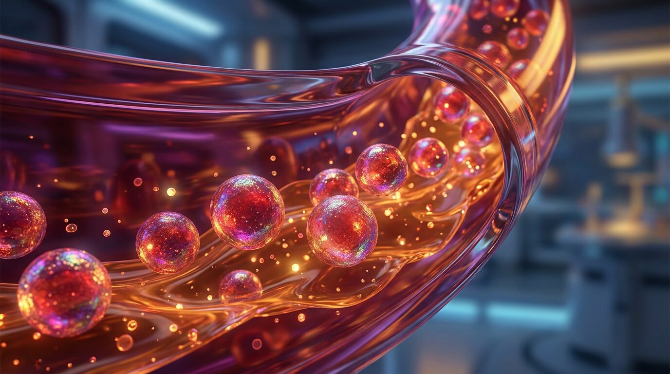
A bag of donated blood is, chemically speaking, a mess. It contains red cells, white cells, platelets, plasma proteins, fragments of cellular machinery, and the quiet biological fingerprints of the donor — blood type, immune markers, trace pathogens screened for but not always absent. Getting that bag from a donor's arm to a trauma patient's vein requires refrigeration, compatibility testing, and a supply chain built on voluntary donation that runs short in disasters precisely when it is needed most. Medicine has been trying to solve this problem for decades, and the closest thing to a real answer looks, at the molecular scale, like a soap bubble filled with hemoglobin.
Hemoglobin vesicles — HbV, in the shorthand of researchers — are exactly what the name suggests: hemoglobin, the oxygen-carrying protein at the center of every red blood cell, packaged inside an artificial lipid membrane. The result is a particle that can pick up oxygen where oxygen is plentiful and release it where oxygen is scarce, mimicking the single most critical function of blood, without any of the living cellular machinery that makes real blood so difficult to store, type, and transport. Research published in Frontiers in Medical Technology[1] by Hiromi Sakai and colleagues lays out what that project actually involves: the chemistry of the membrane layers, the methods for getting hemoglobin stably inside them, and the results of experiments testing whether the system works inside a living body.
The Most Abundant Protein in Your Blood, Miniaturized
As Sakai and colleagues note, hemoglobin is the most abundant protein in blood — a molecule so central to animal life that it shows up, in various forms, across nearly the entire animal kingdom. Inside a red blood cell, it does something elegant and almost counterintuitive: it grabs oxygen loosely enough to release it. The heme group at the heart of each hemoglobin subunit contains an iron atom that binds oxygen in the lungs, where oxygen concentration is high, and lets it go in capillary beds where tissues have been consuming it and the local partial pressure has dropped. It is a reversible transaction, driven by chemistry, happening billions of times per second across your entire circulatory system.
The problem with using raw hemoglobin as a blood substitute — and researchers tried this for years — is that free hemoglobin outside a cell is toxic. Without the protective environment of the red blood cell membrane, it breaks apart, releases its iron in damaging ways, and gets cleared from the bloodstream far too quickly to be clinically useful. The red blood cell is not just a vessel. Its membrane actively manages what goes in and out; its internal chemistry keeps hemoglobin in the right molecular state; its shape maximizes surface area for gas exchange. Strip all of that away and you lose most of what makes hemoglobin work.
The hemoglobin vesicle approach sidesteps this by building a surrogate membrane — a lipid bilayer, chemically similar to the walls of biological cells, assembled around a concentrated payload of purified hemoglobin. The particle that results is smaller than a real red blood cell, which turns out to be useful: it can reach tissue through narrowed or partially obstructed vessels that a full-sized cell might struggle to navigate. The Sakai et al. paper[1] describes the engineering challenges involved in building these structures reproducibly — controlling the thickness and composition of the membrane layers, preventing the hemoglobin inside from oxidizing into methemoglobin (a form that can no longer carry oxygen), and developing preparation methods efficient enough to produce the volumes that clinical use would require.
“Free hemoglobin outside a cell is toxic. The red blood cell is not just a vessel — its membrane actively manages what goes in and out.”
What the Liposome Actually Does
A liposome is a self-assembling structure: phospholipid molecules, which have a water-attracting head and a water-repelling tail, spontaneously organize into a double-layered sphere in an aqueous environment, the same basic geometry as a cell membrane. Researchers have used liposomes as drug-delivery vehicles for years, loading them with chemotherapy agents, antifungals, and other compounds that would be too toxic or too unstable to administer freely — a connection to the broader engineering challenge of getting the right chemistry to the right place inside the body. For HbV, the goal is to capture purified hemoglobin inside that sphere while keeping the lipid shell stable enough to survive storage and circulation.
Getting this right is harder than it sounds. The hemoglobin inside must stay in its oxygen-carrying form — ferrous iron, not oxidized ferric iron — which means the preparation environment has to be carefully controlled for oxygen and reducing conditions. The membrane has to be stable at physiological temperature and pressure, flexible enough to pass through capillaries without rupturing, and chemically unreactive enough not to trigger an immune response. Researchers add a polymer layer — polyethylene glycol is common — to the outside of the vesicle to help it evade immune recognition and extend its circulation time. Each of these is a separate engineering problem with its own failure modes.
The in vivo results described in the research — experiments in animal models — show that HbV can function as an oxygen carrier in living circulation. The particles move through blood vessels, exchange gas, and eventually get cleared by the same immune cells that remove aged red blood cells, without apparent acute toxicity at the doses tested. That is a meaningful result. It is also the kind of result that sits at the early end of a long translational road. Animal models demonstrate biological plausibility. They do not automatically predict what will happen in a human trauma patient receiving HbV under emergency conditions, alongside other interventions, with a compromised circulatory system.
The Shelf-Life Problem and Why It Changes Everything
The practical argument for HbV is not primarily that it is better than donated blood. It is that donated blood, for all its irreplaceable qualities, cannot be everywhere at once. Real red blood cells have a storage life of roughly six weeks under refrigeration, and even within that window, their function degrades. They require type matching — give the wrong ABO or Rh type and the immune system attacks the transfused cells with sometimes fatal results. In a mass-casualty event, a remote trauma scenario, a military field setting, or a low-resource healthcare environment, the cold chain and compatibility infrastructure that make blood transfusion safe in a hospital simply may not exist.
Hemoglobin vesicles, because they carry no cell-surface antigens, would in principle be universally compatible — no blood typing required. And because they are not living cells, they can potentially be stabilized for much longer storage, possibly at room temperature in a lyophilized (freeze-dried) form that reconstitutes with water. The Sakai research program describes this storable, ready-to-use formulation as a central goal. If it works, the clinical vision is something like a sealed unit that sits in an ambulance, a field medic's kit, or a rural clinic for months, then gets used in the first critical minutes of hemorrhagic shock before a hospital is reachable. That is the gap in trauma care that kills people — not the absence of a perfect blood substitute, but the absence of any oxygen carrier at all in the first hour.
“Hemoglobin vesicles carry no cell-surface antigens — in principle, no blood typing required.”
What HbV Cannot Do
It is worth being precise about what hemoglobin vesicles are not attempting to replace. Blood is not only an oxygen delivery system. It is a transport network for nutrients, hormones, immune cells, and waste products. It is a clotting system — platelets and the cascade of coagulation proteins that stop bleeding when a vessel is damaged, a process as intricate as any emergency repair the body undertakes. It is an immune organ, carrying the white cells that surveil tissue for infection and cellular damage. A hemoglobin vesicle does none of these things. It delivers oxygen. That is the one function it is built for, and in the specific context of hemorrhagic shock — where the immediate threat to life is tissue hypoxia from blood loss — that one function may be enough to buy the time a patient needs.
The research also does not yet resolve questions about what happens in repeated or large-volume HbV administration, whether there are inflammatory responses that emerge at clinical doses, or how the particles interact with the compromised physiology of a trauma patient rather than a healthy research animal. These are not objections to the research — they are the next set of questions it has to answer. Translational medicine is a process of narrowing uncertainty, not eliminating it, and HbV sits at an early point on that path.
The Hidden Engineering in a Bubble of Fat
There is something quietly remarkable about the hemoglobin vesicle as an object. It is, at its core, a piece of biological mimicry: researchers taking the functional principle of a red blood cell — iron-centered oxygen binding inside a lipid envelope — and rebuilding it from purified components, outside the context of living biology, in a form that can be manufactured, stored, and shipped. The lipid membrane is not alive. The hemoglobin inside is purified protein. Yet together they perform the essential chemistry that keeps tissue alive when blood is gone.
This is what makes the hemoglobin vesicle project interesting not just as a medical technology but as a demonstration of how deeply we have come to understand the mechanisms of ordinary physiology. We know enough about how a red blood cell works — at the level of membrane architecture, protein chemistry, and gas exchange physics — to begin rebuilding its core function from scratch. The resulting particle is simpler than a cell by orders of magnitude. It cannot adapt, reproduce, or signal. But it can do the one thing that matters most when a person is bleeding out in the back of an ambulance, far from a blood bank, with a medic and a few minutes: carry oxygen to the tissue that is running out of it.
References
- Research of storable and ready-to-use artificial red blood cells (hemoglobin vesicles) for emergency medicine and other clinical applications - PubMed (pubmed.ncbi.nlm.nih.gov)
Describes hemoglobin vesicle engineering challenges, membrane composition, oxidation prevention, and in vivo animal model results demonstrating oxygen-carrying function.
About Aris Thorne
Aris Thorne is a microbiologist who writes about the hidden mechanics of ordinary life: the microbes running your home, the chemistry unfolding in food and water, the physics built into familiar objects, and the biological systems quietly keeping the human body alive. His work follows science from kitchens, bathrooms, dust, soil, and city air into wounds, immune responses, infections, medicines, cells, and other worlds. He is most interested in the moment something familiar stops looking simple and reveals the living machinery underneath.
More like this
Your Bruise Is Changing Color for the Same Reason Leaves Do
That purple mark spreading under your skin isn't damage lingering — it's a molecular demolition crew working through a precise, color-coded sequence to erase the evidence.
Your Brain Now Contains a Spoonful of Plastic. We Don't Know What It's Doing.
A landmark study found microplastics concentrating in human brain tissue at startling levels — and the question of what they're doing there may be the most important unanswered problem in modern medicine.
The Slime You Scrubbed Off the Drain Came Back. That's the Point.
New research shows that household cleaning kills the bacteria inside a biofilm while leaving its scaffold intact — which means the colony doesn't die, it just takes a short nap.