Your Bruise Is Changing Color for the Same Reason Leaves Do
That purple mark spreading under your skin isn't damage lingering — it's a molecular demolition crew working through a precise, color-coded sequence to erase the evidence.
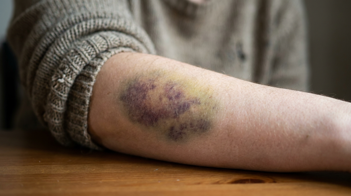
Press a finger against a fresh bruise and you will feel it before you see much: a tenderness that seems to pulse slightly, sitting just under the surface like something is still happening down there. Which is exactly right. A bruise is not an injury in the way a cut is an injury. A cut is an opening. A bruise is a closed system — a sealed chamber where blood has leaked into tissue it was never meant to occupy, and where your body has already begun one of its more elaborate clean-up operations.
The color is the part most people notice without really thinking about. That first dark red or purple. Then the shift toward blue, toward green, toward yellow, and finally nothing — skin returning to normal over a week or two as if the whole event quietly resolved itself. It looks like fading. It is not fading. It is a sequence of chemical reactions moving through your tissue in a specific order, driven by enzymes, broken down into molecular fragments small enough to carry away in your bloodstream, each stage of demolition announcing itself through a different color. The bruise is not healing by becoming less. It is healing by becoming something else.
The comparison to autumn leaves is not poetic license. It is the same underlying chemistry — hemoglobin and chlorophyll are both large pigment molecules built around a metallic core, and when biological systems break them down, they produce similar intermediate compounds that move through the visible spectrum as their molecular structure changes. Leaves lose their chlorophyll and reveal the pigments underneath. A bruise loses its hemoglobin and converts it, step by step, into smaller and smaller molecules, each one absorbing light differently. The tree and the bruise are running almost the same enzymatic program.
Understanding why requires going back to the moment of impact — to the split second when something strikes your body hard enough to rupture the small blood vessels just beneath your skin.
The Rupture
Your skin has no trouble surviving the force that causes a bruise. It is the capillaries and small venules underneath — the finest branches of your circulatory system — that give way. These vessels are extraordinarily thin-walled, some of them just a single cell thick, and a sharp enough impact shears them open. Blood escapes into the interstitial space: the soft connective tissue between cells that is not built to hold it. Because the skin remains intact above, that blood has nowhere to go. It pools.
The first color you see, that deep red-purple, is simply the color of deoxygenated blood seen through a few millimeters of skin and fat. Hemoglobin — the large protein inside red blood cells that carries oxygen through your arteries — appears bright red when it is oxygenated, and dark, slightly purple-red when it is not. Blood pooled in tissue is cut off from oxygen replenishment, so it shifts toward the deoxygenated end of the spectrum almost immediately. The bruise looks dark because the blood inside it is dark, and it is dark because it is stranded.
Meanwhile, your immune system has already detected the problem. Mast cells in the surrounding tissue release histamine, which dilates nearby blood vessels and makes them more permeable. This is why bruised tissue swells and feels warm — it is being deliberately flooded with immune cells, particularly macrophages, which are about to do most of the actual work. The initial redness and warmth that accompany a bruise are not signs of something going wrong. They are the body opening its operational channels.
What Macrophages Actually Do
The word macrophage comes from the Greek for large eater, which is fairly accurate. These are large, mobile immune cells whose primary function is phagocytosis — engulfing cellular debris, pathogens, and foreign material and digesting it internally. When blood pools in tissue, the red blood cells within it begin to break down. Their membranes deteriorate. Hemoglobin leaks out. And macrophages move in to consume it.
“The bruise is not healing by becoming less. It is healing by becoming something else.”
Inside the macrophage, hemoglobin encounters an enzyme called heme oxygenase[2]. This enzyme cleaves the heme group — the iron-containing ring at the center of hemoglobin — away from the surrounding protein. The protein portion gets broken into amino acids and recycled. The heme ring itself is then cracked open, and what emerges is a straight-chain compound called biliverdin. Biliverdin is green. This is where the bruise turns green, typically around days three through five, and it is a direct readout of heme oxygenase doing its job.
Biliverdin does not stay green for long. Another enzyme, biliverdin reductase, converts it into bilirubin — a yellow-orange compound that is far more water-soluble than its predecessor. This matters because water-soluble molecules can be transported. Bilirubin gets picked up by albumin in the blood, carried to the liver, conjugated with glucuronic acid to make it even more soluble, and eventually excreted in bile. The yellow stage of a bruise is bilirubin becoming mobile enough to leave. This is the body finishing its cleanup, loading the last of the molecular debris onto trucks.
The iron that was released when heme was cleaved is handled separately. Your body is intensely conservative with iron — it is too metabolically expensive to waste. Macrophages store it inside a protein called ferritin, then release it back into circulation in controlled amounts, where it gets reused to build new hemoglobin in fresh red blood cells. Nothing about this process is casual. The body runs an extraordinarily precise iron economy, and a bruise is one of the places where that economy becomes briefly visible.
Why the Colors Move the Way They Do
Most bruises do not change color uniformly. The edges tend to green and yellow before the center does. This is not random. The perimeter of the pooled blood is where macrophage activity is densest, because that is where circulating immune cells encounter the blood most readily — from the outside in. The center of the bruise, particularly in a large one, is the most metabolically isolated. Macrophages are working their way inward. The color gradient you see is a spatial map of enzymatic progress.
Bruises also frequently appear to migrate over time, sliding slightly downward from their original site. This is gravity doing what gravity does. Bilirubin and other breakdown products are water-soluble enough to move through the interstitial fluid, and they will drift along the path of least resistance, pooling in looser connective tissue slightly below where the original impact occurred. A bruise on your thigh might seem to travel toward your knee. The injury has not moved. The chemical residue of it has.
“The yellow stage of a bruise is bilirubin becoming mobile enough to leave — the body loading the last of the molecular debris onto trucks.”
Certain areas of the body bruise more visibly because the skin there is thinner and the subcutaneous fat is minimal, making pooled blood easier to see from the surface. The backs of elderly people's hands are a classic example — the skin thins with age as collagen production slows, and vessels that might have held under the same impact in a younger person give way more easily. Bruising more easily as you age is not primarily about fragile blood — it is about fragile scaffolding around the vessels.
The Autumn Leaf Problem
Chlorophyll and hemoglobin are not the same molecule, but they belong to the same structural family: both are built around a porphyrin ring, a flat, nitrogen-rich molecular scaffold with a metal atom at its center. In hemoglobin, that metal is iron. In chlorophyll, it is magnesium. And both molecules are degraded by similar enzymatic pathways that crack open that central ring and produce chain-like breakdown products that absorb light in progressively shifting ways.
When a tree pulls resources back from its leaves in autumn, chlorophyll breaks down. As the green pigment disappears, the yellows and oranges of carotenoids — which were present in the leaf all along, masked by chlorophyll's intensity — become visible. The reds and purples in some species come from anthocyanins, which are actively synthesized during senescence. The leaf is not dying into color. It is undergoing a structured molecular dismantlement that happens to be beautiful.
A bruise runs a parallel script. The red-purple of hemoglobin gives way to the green of biliverdin, then the yellow of bilirubin — not because the colors were hidden underneath waiting to be revealed, but because each color is a different molecule produced in sequence by enzymatic activity. The tree and the bruise are not merely analogous. They are both visible evidence of porphyrin chemistry moving through its breakdown stages, rendered legible by the way pigment molecules absorb different wavelengths of light as their ring structures are cleaved and modified.
When the System Signals Something Else
The normal bruise resolves in one to three weeks depending on size, depth, and location. But the same chemistry that makes bruising comprehensible also makes certain deviations from it medically meaningful. Bilirubin is normally processed by the liver quickly enough that it never accumulates visibly in tissue. When the liver is overwhelmed — by illness, by hemolytic conditions where red blood cells are breaking down faster than normal, or by bile duct obstruction — bilirubin backs up into the bloodstream and diffuses into tissue throughout the body. The result is jaundice: skin and whites of the eyes turning yellow, the same yellow as the late-stage bruise, because the same molecule is involved.
Spontaneous bruising — bruises that appear without any impact the person can recall — can indicate problems with platelet function or clotting factor deficiency, since platelets and clotting proteins are normally the first responders to vascular damage, forming an initial plug that limits how much blood escapes before repair begins. If that primary hemostasis is compromised, even trivial trauma allows enough blood to pool to form a visible bruise. The bruise itself is always the same chemistry. What changes is the threshold that triggers it.
There are also conditions in which heme oxygenase is deliberately upregulated — meaning the body runs its heme-dismantling process faster and more aggressively than normal. This happens in certain inflammatory states and in response to oxidative stress. Biliverdin and bilirubin are not merely waste products; research into heme oxygenase activity has found that both compounds have antioxidant properties[1], and the enzyme pathway that produces them appears to function partly as a cellular stress response. The bruise-color chemistry is also, in a quieter register, part of how your cells manage damage at the molecular level.
What the Color Is Telling You
“The tree and the bruise are both visible evidence of porphyrin chemistry moving through its breakdown stages, rendered legible by the way pigment molecules absorb light differently as their ring structures are cleaved.”
There is something almost legible about a bruise, once you know the code. The dark purple is stranded, deoxygenated blood — the raw material of the problem. The blue is that same blood deepening in the tissue, oxygen long gone. The green is heme oxygenase at work, cracking the porphyrin ring, converting iron-rich heme into biliverdin. The yellow is the exit signal — bilirubin going water-soluble, entering the circulatory system, heading toward the liver and eventually out of the body entirely. Each color is a stage report from a process so organized it would be impressive if it were designed rather than evolved.
It was evolved, of course — over hundreds of millions of years, in organisms that needed efficient ways to manage vascular damage without losing precious iron or letting degraded red blood cells trigger unchecked inflammation. The same pathway exists in fish, in amphibians, in birds. Heme oxygenase is one of the most conserved enzymes in vertebrate biology[3], which is a way of saying that evolution found this solution early and kept it. Every bruise you have ever had has been running ancient software, written into your biology long before anything remotely like you existed.
Next time you catch a bruise turning that unmistakable yellow at the edges — that color that looks almost sickly, almost wrong — it is worth pausing on what you are actually seeing. Not injury persisting. Not your body failing to recover fast enough. You are watching bilirubin becoming transportable, watching your macrophages finish their work, watching iron get inventoried and filed away for future use. The yellow means the job is nearly done. The bruise is not fading. It is leaving.
References
- Biliverdin reductase: A major physiologic cytoprotectant (pnas.org)
Establishes that biliverdin reductase and its products have antioxidant properties during bruise breakdown. - Biochemistry, Biliverdin (ncbi.nlm.nih.gov)
Explains that heme oxygenase breaks hemoglobin into biliverdin and bilirubin, the green and yellow compounds visible in bruise color progression. - Heme Oxygenase-1, Oxidation, Inflammation, and Atherosclerosis (frontiersin.org)
- Porphyrins One Ring In The Colors Of Life (americanscientist.org)
Provides the structural definition of porphyrin rings, the metal-centered molecular scaffold shared by hemoglobin and chlorophyll.
About Aris Thorne
Aris Thorne is a microbiologist who writes about the hidden mechanics of ordinary life: the microbes running your home, the chemistry unfolding in food and water, the physics built into familiar objects, and the biological systems quietly keeping the human body alive. His work follows science from kitchens, bathrooms, dust, soil, and city air into wounds, immune responses, infections, medicines, cells, and other worlds. He is most interested in the moment something familiar stops looking simple and reveals the living machinery underneath.
More like this
Scientists Are Building Artificial Blood From the Inside Out
Hemoglobin vesicles can carry oxygen without a single living cell — Deoxy HbV — and they might one day sit on a shelf in an ambulance, waiting for you.
Why a Tiny Cut Can Trigger Your Immune System to Attack Itself
Infection isn't just invasion — it's a molecular negotiation where pathogens rewrite your cells' operating instructions and the body's defenses sometimes make things catastrophically worse.
Your Brain Has a Drain, and It Only Opens When You're Asleep
While you're unconscious, a hidden plumbing system floods your brain with fluid and hauls away the toxic waste that builds up while you think — and scientists are only beginning to understand what happens when it can't.