Your Brain Has a Drain, and It Only Opens When You're Asleep
While you're unconscious, a hidden plumbing system floods your brain with fluid and hauls away the toxic waste that builds up while you think — and scientists are only beginning to understand what happens when it can't.
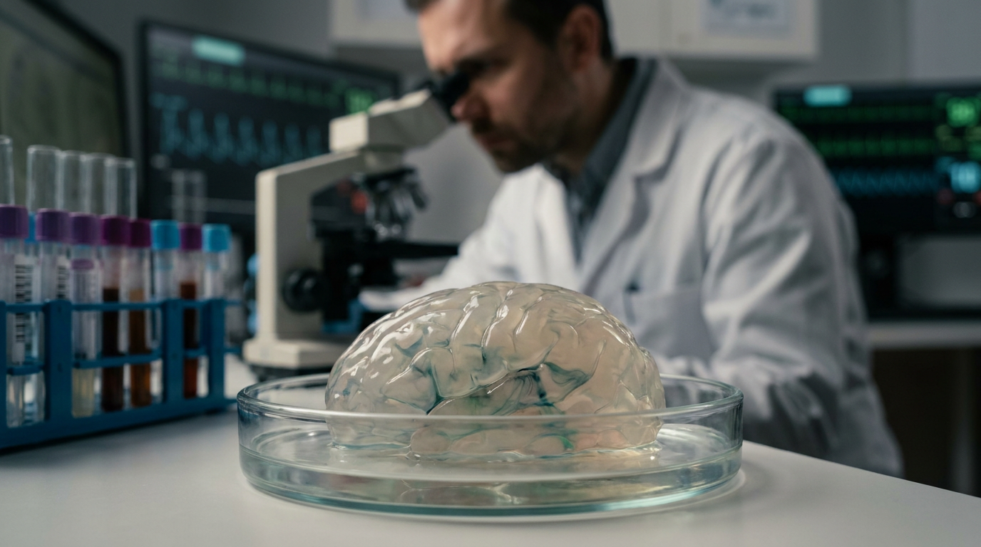
Sometime around two in the morning, while you are completely unaware of anything, your brain does something it cannot do while you are awake. The spaces between your neurons — narrow corridors called the interstitial space, collectively forming a kind of spongy inner landscape — expand. Cerebrospinal fluid, the clear liquid that normally bathes the brain's outer surface, begins moving through those corridors with unusual purpose, driven in pulses that follow the rhythm of your heartbeat and your slow, sleeping breath. It floods in. It carries things out. And then, in the hours before you wake, it recedes, leaving the tissue quieter, cleared, reset.
This is the glymphatic system, and it is one of the more surprising discoveries neuroscience has made in the last fifteen years. The name blends glial and lymphatic: the brain lacks the lymphatic vessels that drain waste from the rest of the body, but its glial cells — particularly the star-shaped astrocytes that wrap around blood vessels — have built something that works on a similar principle. Channels made from proteins called aquaporin-4 line the astrocyte endfeet[1] pressed against blood vessel walls, and through these channels, cerebrospinal fluid can move into the brain tissue itself, washing through the interstitial space and eventually draining out through pathways along cranial nerves into the lymphatic vessels of the neck. It is a plumbing system hidden inside an organ that scientists spent more than a century assuming had no plumbing at all.
The reason no one found it earlier is partly a matter of timing. The glymphatic system does most of its work when you are asleep, and studying a sleeping brain is harder than studying a waking one. The infrastructure itself is also subtle — it operates through existing anatomical channels rather than dedicated new vessels, and it moves fluid slowly, by the standards of the circulatory system, driven by pressure gradients and the pulsing of arteries rather than a dedicated pump. Once researchers started looking at living animals during sleep using two-photon microscopy and fluorescent tracers, the system came into view with startling clarity: fluid moving through channels, metabolic waste accumulating, the brain doing what amounts to its nightly dishwashing.
What the brain is washing away matters enormously. Neurons are metabolically expensive — they burn glucose at a high rate and generate byproducts continuously. Among those byproducts are proteins that, in excess, become dangerous: amyloid-beta, which tends to misfold and clump into plaques, and tau, which can tangle into structures that strangle cellular transport. Both are strongly linked to Alzheimer's disease. And both, studies in animal models have shown, are cleared from the brain significantly faster during sleep than during wakefulness. The glymphatic system appears to be, among other things, the mechanism by which the brain takes out the molecular trash it generates just by being alive and thinking.
The Brain's Hidden Hydraulics
To understand how the system actually moves fluid, it helps to picture the brain's vascular architecture. Arteries enter the brain and branch into smaller vessels, each one wrapped in a sleeve of astrocyte endfeet that forms a space called the perivascular space. Cerebrospinal fluid, produced by specialized cells in the choroid plexus of the brain's ventricles, flows into these perivascular spaces along arteries and then passes through the aquaporin-4 channels into the surrounding interstitial space — the narrow, fluid-filled gaps between cells where metabolic waste collects. From there, the fluid exits along veins, draining toward the cervical lymph nodes and eventually into general circulation.
What drives this flow is not, as you might expect, a dedicated pump. The primary engine appears to be arterial pulsation — the rhythmic expansion and contraction of blood vessels with each heartbeat creates pressure waves that push cerebrospinal fluid through the perivascular spaces. Respiration adds another layer of drive, with the pressure changes of each breath modulating flow direction and rate. During slow-wave sleep, when heartbeat and breathing fall into slow, synchronized rhythms, these pressure waves become more regular and effective. The interstitial space itself physically expands during sleep — by roughly sixty percent in some studies of mouse brains[2] — which lowers resistance and lets the fluid move more freely through tissue that was, minutes ago, packed too tightly to permit the same flow.
“The interstitial space expands by roughly sixty percent during sleep, opening corridors through brain tissue that are simply too narrow to use while you are awake.”
The expansion of the interstitial space is itself regulated by norepinephrine, a neurotransmitter that stays elevated during wakefulness and drops sharply during sleep. Norepinephrine causes cells to take in water and swell slightly, which compresses the interstitial space and limits glymphatic flow. Remove norepinephrine signaling — as happens naturally when you fall asleep — and the cells relax, the spaces open, and the plumbing works. This means the glymphatic system is not simply dormant during the day. It is actively suppressed, held in check by the same arousal chemistry that keeps you alert and functional. Sleep doesn't just allow the system to work. It is a prerequisite for it.
Amyloid Clocks and the Cost of Staying Awake
Amyloid-beta is produced continuously in the brain, a byproduct of normal synaptic activity. In a healthy brain that sleeps adequately, it is cleared fast enough that it never accumulates to dangerous concentrations. In a brain that is chronically sleep-deprived, clearance falls behind production, and the protein begins to aggregate — first as soluble oligomers that interfere with synaptic function, then eventually as the dense plaques visible in Alzheimer's pathology. Studies using PET imaging in human volunteers[3] found that even a single night of sleep deprivation produced a measurable increase in amyloid-beta accumulation in regions including the hippocampus and thalamus, areas critical to memory and sensory processing. The increase was not catastrophic in a single night — the brain is not a machine that breaks instantly — but it demonstrated that amyloid clearance is not a slow background process. It is time-sensitive in a way tied directly to sleep cycles.
Tau behaves similarly. During wakefulness and especially during sleep deprivation, tau concentrations in the cerebrospinal fluid rise. Tau normally stabilizes the internal scaffolding of neurons — the microtubules that give cells their shape and serve as tracks for molecular transport. When tau misfolds and aggregates, that scaffolding fails, and neurons begin to lose their structural integrity. In Alzheimer's disease, tau tangles spread through the brain in a pattern that corresponds closely to the progression of cognitive decline. The connection between poor sleep and tau accumulation is not yet fully mechanistic in humans — research is ongoing — but the directional evidence is consistent enough that sleep disruption is now considered a risk factor for Alzheimer's development rather than merely a symptom of it.
“Sleep disruption is now considered a risk factor for Alzheimer's development, not merely a symptom — a shift that quietly changes how researchers think about the disease's timeline.”
Why Position Matters
One of the stranger findings to emerge from glymphatic research is that body position during sleep appears to affect how efficiently the system clears waste. Studies in rodents found that sleeping on the side[4] — the lateral position — produced more efficient glymphatic transport than sleeping on the back or stomach. The proposed mechanism involves the geometry of perivascular drainage: in lateral sleep, gravity and the arrangement of the brain's drainage pathways may combine more favorably, reducing the distance and resistance that cerebrospinal fluid must overcome. Humans, in practice, tend to sleep on their sides more than any other position, a behavioral pattern that may be quietly adaptive in ways we did not previously recognize.
The posture finding has not yet been confirmed in large human trials with the same rigor as the rodent work — the difficulty of controlling sleep position across hundreds of subjects, combined with the complexity of measuring glymphatic flow in living humans, makes this an active area of investigation rather than settled science. But it illustrates something important about glymphatic research as a field: the system has properties that feel almost designed to be counterintuitive. The idea that how you orient your body during unconsciousness influences the chemistry of waste clearance in your brain is not obviously suggested by anything in the experience of going to sleep. It is exactly the kind of mechanism that stays hidden until someone builds the tools to look for it.
Sleep Stage, Not Just Sleep Duration
Not all sleep contributes equally to glymphatic function. Slow-wave sleep — the deep, non-REM stage characterized by large, synchronized neural oscillations called slow waves — appears to be the phase in which glymphatic clearance operates most intensively. During slow-wave sleep, neuronal activity is highly coordinated: large populations of neurons fire together, then rest together, producing the rhythmic electrical signature that gives this stage its name. These oscillations are accompanied by coordinated fluctuations in blood flow and cerebrospinal fluid movement that appear to amplify glymphatic drive. The slow wave is, in this sense, not just a feature of deep sleep but something closer to a mechanism for it — the brain synchronizing its activity in a way that turns hydraulic efficiency to maximum.
REM sleep, the stage associated with vivid dreaming and rapid eye movement, plays a different role. Brain activity during REM is high and somewhat similar to waking patterns, and glymphatic flow appears to be lower in this stage. This does not mean REM is without biological purpose — it is essential for emotional memory consolidation and other processes — but the specific work of metabolic waste clearance seems concentrated in the slow-wave periods that dominate the earlier part of the night. This creates an uncomfortable implication for modern sleep habits: the sleep many people sacrifice first, by staying up late or drinking alcohol that suppresses slow-wave sleep, is disproportionately the sleep that runs the brain's waste-disposal system.
Alcohol is worth pausing on. It is often experienced as a sleep aid — it reduces the time to fall asleep and produces early sedation — but it fragments sleep architecture significantly, suppressing slow-wave sleep and increasing waking during the second half of the night. The subjective experience is of easier sleep onset with worse sleep quality overall. For glymphatic function, this is approximately the opposite of helpful: sedation is not the same as the organized, oscillating deep sleep that drives waste clearance, and a brain that spends less time in slow-wave sleep accumulates more of the metabolic byproducts that the system exists to remove.
Aging, Dysfunction, and the System's Slow Failure
Glymphatic function declines with age, and the mechanisms of that decline point toward several overlapping failures. The aquaporin-4 water channels in astrocyte endfeet lose their organized arrangement — a process called depolarization — which reduces the efficiency of fluid transport through brain tissue. The slow-wave sleep that drives clearance becomes shorter and shallower with age, further reducing the hydraulic drive that the system depends on. Blood vessel stiffness increases, which dampens the arterial pulsations that push cerebrospinal fluid through perivascular spaces. Each of these changes is individually modest; together, they compound into a system running at a fraction of its youthful capacity.
The timing of this decline is grimly suggestive. Amyloid-beta begins accumulating in the brain years — sometimes decades — before the cognitive symptoms of Alzheimer's disease appear. Tau pathology follows a similar slow arc. The window in which glymphatic dysfunction might allow these proteins to accumulate without yet causing detectable symptoms corresponds closely to the period in which interventions might theoretically be most effective. This is part of why researchers in the field take the glymphatic system seriously as a therapeutic target: not because clearing waste during sleep is an exotic luxury, but because it may be among the most time-critical biological processes the aging brain depends on.
“The window in which glymphatic dysfunction allows toxic proteins to accumulate without yet causing symptoms may be exactly when intervention could matter most.”
Current research is exploring several routes to support or restore glymphatic function: compounds that modulate aquaporin-4 expression, approaches to enhance slow-wave sleep quality in older adults, and even physical interventions like exercise, which appears to improve both sleep architecture and glymphatic clearance in animal models. None of these is a therapy yet in any clinical sense, and the gap between a promising mechanism in rodents and a proven human intervention is a wide one that the history of neuroscience has crossed only slowly. But the conceptual shift is already real: sleep is not the passive absence of consciousness that it resembled from the outside for so long. It is a phase of active biological maintenance, timed and organized and necessary in ways that waking life cannot substitute for.
What It Means to Lie Down
There is something quietly vertiginous about knowing this. Every night you fall asleep, your brain shifts into a mode your waking self has no access to and no awareness of: fluid moving through tissue, channels opening, proteins being swept out of the spaces between neurons that spent the day thinking, remembering, worrying, and noticing. The system is not dramatic. It does not announce itself. It runs on the slow architecture of sleep — on the expanding of cellular corridors and the rhythm of arteries and the long, synchronized oscillations of neurons resting together — and it clears what the day left behind in the only window it gets. The waste that accumulates during consciousness, including the proteins most associated with the brain's most feared disease, waits for this. It has no other mechanism to move it. That is what you are doing when you sleep. Not nothing. The opposite of nothing.
References
- A Paravascular Pathway Facilitates CSF Flow Through the Brain Parenchyma and the Clearance of Interstitial Solutes, Including Amyloid β (science.org)
Identifies aquaporin-4 protein channels in astrocyte endfeet as the structural basis for cerebrospinal fluid movement into brain tissue. - Sleep Drives Metabolite Clearance from the Adult Brain (science.org)
Provides the quantitative finding that interstitial space expands roughly sixty percent during sleep in mouse brains, enabling glymphatic flow. - β-Amyloid accumulation in the human brain after one night of sleep deprivation (pnas.org)
Demonstrates via PET imaging that a single night of sleep deprivation increases amyloid-beta accumulation in human brain regions including hippocampus and thalamus. - The Effect of Body Posture on Brain Glymphatic Transport (jneurosci.org)
Shows that lateral (side) sleeping position produces more efficient glymphatic transport than back or stomach positions in rodent studies.
About Aris Thorne
Aris Thorne is a microbiologist who writes about the hidden mechanics of ordinary life: the microbes running your home, the chemistry unfolding in food and water, the physics built into familiar objects, and the biological systems quietly keeping the human body alive. His work follows science from kitchens, bathrooms, dust, soil, and city air into wounds, immune responses, infections, medicines, cells, and other worlds. He is most interested in the moment something familiar stops looking simple and reveals the living machinery underneath.
More like this
Your Brain Washes Itself at Night. Here's What Breaks the Drain.
A 2025 trial confirmed the brain flushes Alzheimer's proteins overnight — but the specific sleep conditions that make that flush work are messier, and more contested, than anyone is telling you.
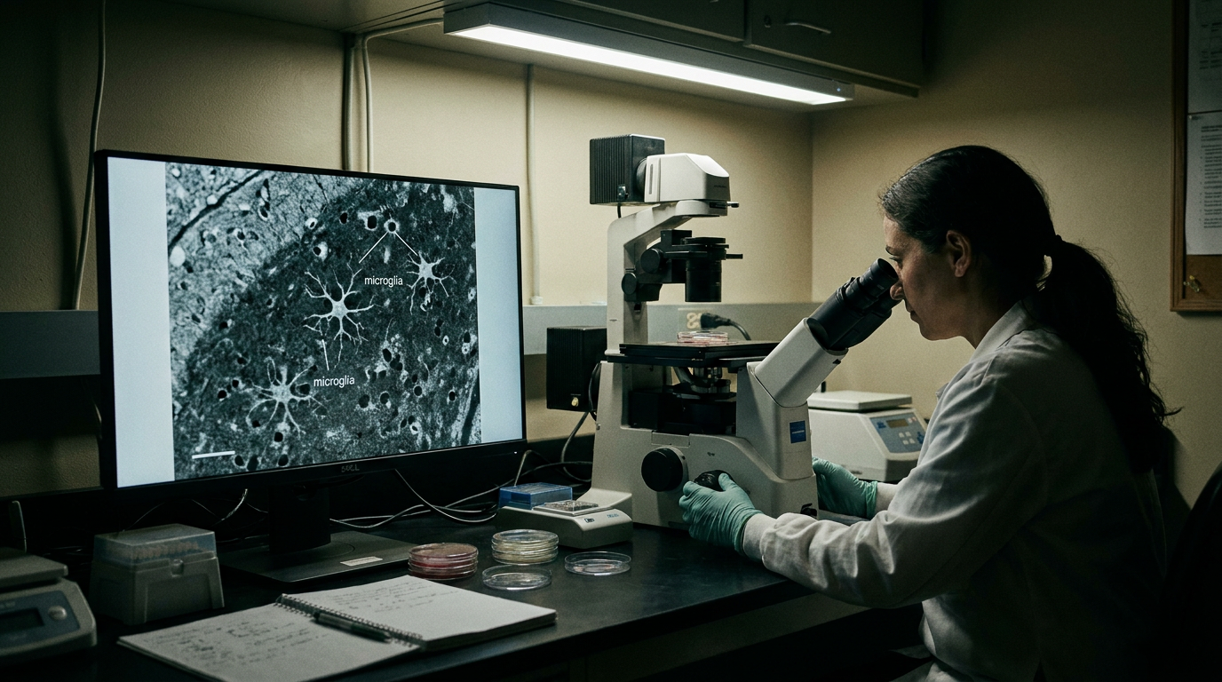
The Brain's Immune System Is Supposed to Protect You. With Age, It Starts Working Against You.
Microglia — the brain's resident immune cells — spend a lifetime learning to protect you, then gradually flip into a state that erodes the very circuits they were built to defend.

Your Body Clock Is a Better Dementia Predictor Than Your Sleep Hours
A major study found that people with fragmented circadian rhythms had 2.4 times the dementia risk — and the mechanism points to something sleep duration alone can't fix.